Accessory Breast Removal Recovery: Timeline, Scars & What to Expect
Understanding Accessory Breast Removal recovery process, timeline, scar management, compression & activity guidelines can help patients prepare for the surgery.
April 5, 2026


Public parking available or drop off along Arab Street, Village Hotel Bugis (Previously known as Golden Landmark Hotel)
Buses (2, 12, 32, 33, 130, 133, 133A, 520, 960, NR7) MRT (alight at Bugis MRT station)
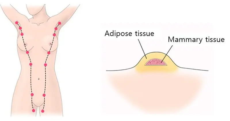
Accessory breast tissue (polymastia) affects 2–6% of women and 1–3% of men, developing along the embryonic milk line and most commonly appearing in the axillary (armpit) area.
The procedure may be covered by insurance and Medisave when deemed medically necessary.
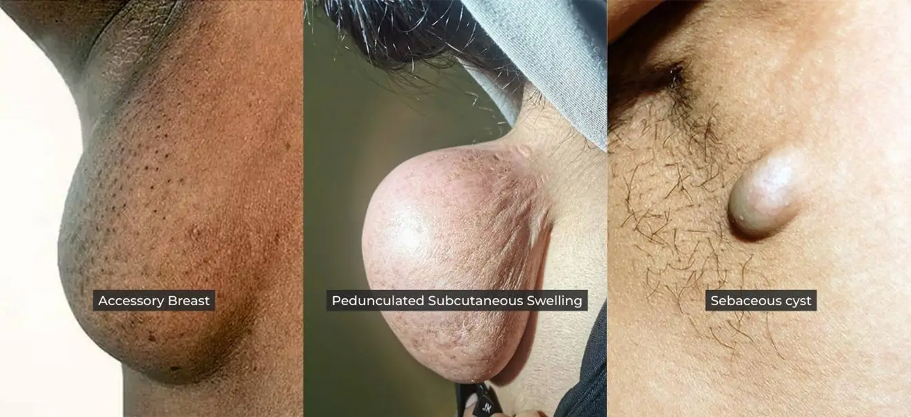
Unlike armpit fat, accessory breast tissue is glandular in nature. This extra tissue creates a noticeable bulge in the armpit and may become swollen or painful during hormonal changes, such as during pregnancy or menstruation. It may also restrict movement, cause skin irritation, and lactate during breastfeeding.
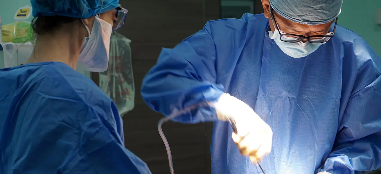
Diagnosis typically involves a clinical examination. At Amaris B. Clinic, Dr Ivan Puah uses a Glandular-Specific Excision Protocol to remove tissue precisely, with minimal scarring and results that are designed to be permanent.
Book a consultation with Dr Ivan Puah at Amaris B. Clinic in Singapore today.
Symptoms of the condition include:
Some pregnant women with this medical condition experience milk secretion. The irritation experienced from clothing may also indicate the presence of accessory breast tissue.


| Treatments | Armpit Fat | Accessory Breast |
|---|---|---|
| Fat | ✓ | There may be presence of armpit fat as well as accessory breast tissue at the axillary area. |
| Glandular tissue | X | ✓ |
| Pain | X | Pain, tenderness and soreness |
| Mass in armpit area | Soft fatty lump | Firm |
| Respond to hormonal fluctuation | X | May feel sore during puberty, menstruation, pregnancy, breastfeeding. May also lactate during breastfeeding. |
| Looks like normal breast | X | Depends on severity |
| Localised to armpit | ✓ | May grow anywhere along the embryonic milk line |
According to Kajava, there are EIGHT grades of the condition[4]. Grade IV is the most common.
Consists of a complete breast with a nipple, areola and glandular tissue.
Consists of glandular tissue and nipple without areola.
Consists of glandular tissue and areola without a nipple.
Consists of glandular tissue only.
Consists of only nipple and areola without glandular tissue.
Consists of only the nipple.
Consists of only the areola.
Consists of only hair.
Healthcare professionals have classified supernumerary nipples according to their size, shape and tissue components. The primary types include:
| Types | Glandular tissue | Nipple | Areola | Fat tissue | Hair patch |
|---|---|---|---|---|---|
| Polymastia (supernumerary breasts) | ✓ | ✓ | ✓ | ✓ | |
| Supernumerary nipple | ✓ | ✓ | |||
| Supernumerary nipple | ✓ | ✓ | |||
| Aberrant glandular tissue | ✓ | ||||
| Pseudomamma | ✓ | ✓ | ✓ | ||
| Polythelia | ✓ | ||||
| Polythelia areolaris | ✓ | ||||
| Polythelia pilosa | ✓ |
Several factors are considered when evaluating candidates for accessory breast removal surgery.
You should not be pregnant, breastfeeding or have any underlying medical condition.
Smoking and alcohol will hinder post-op recovery and results.
It is important to have realistic expectations and achievable results based on your clinical condition.
Diagnosis of accessory breast tissue involves identifying the existence of extra axillary glandular tissue.
The doctor will examine the armpit area to look for a soft-tissue mass or thickening.
Ultrasound results can show the tissue beneath, which will look the same as a normal breast glandular tissue.
OR
Mammography can be used to assess extra tissue with a more detailed view, including the imagery of the axillary tail of Spence.
During the surgery, biopsies of the excised tissue will be sent for histology to determine the nature of the tissue.
Unlike generic methods, at Amaris B. Clinic, we leverage three diagnostic pillars to guide treatment:


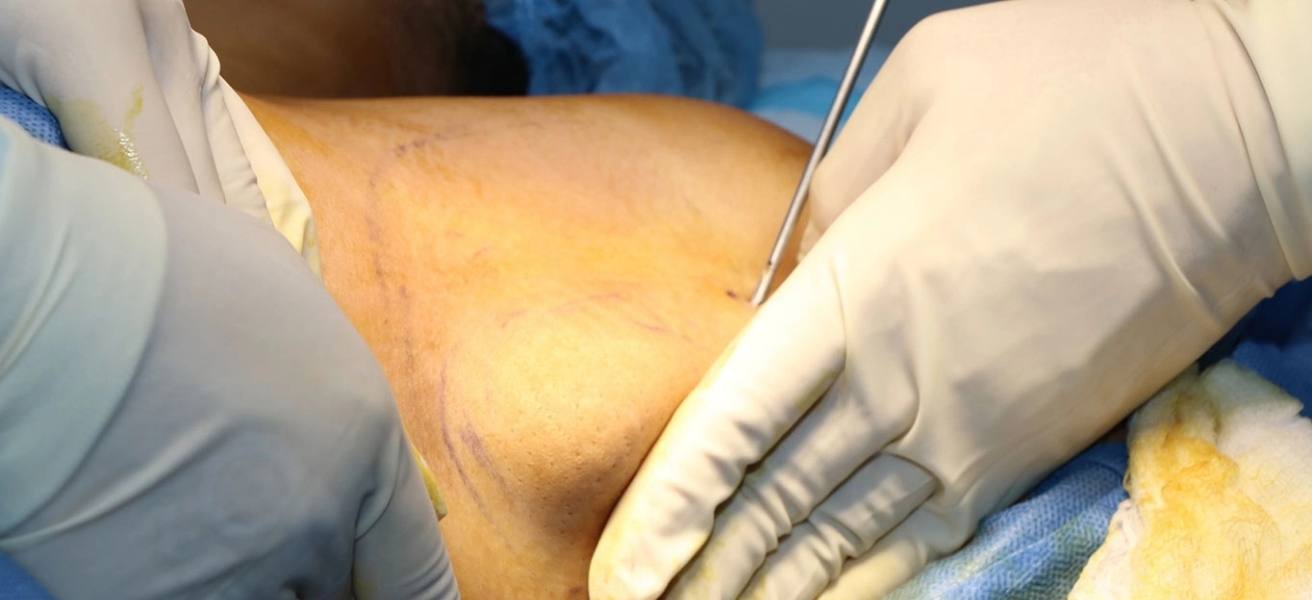
Hidden 3 to 4 cm incision within the natural axillary crease (virtually undetectable post-recovery).
Infiltrate anaesthetic fluid to the targeted area to:
Specialised dissecting instruments remove tissue layer-by-layer.
Tension-adjusted suturing with 4-0 subcuticular stitch for scarless surface healing.
Dressings applied.
Excised tissue sent for pathological examination.
Say goodbye to the bulge for a smoother and more contoured appearance.
Helps relieve discomfort or pain that excess tissue may cause.
Many individuals feel more self-assured and willing to participate in social activities.
Reduces feelings of self-consciousness, leading to a happier, more fulfilling life.
Accessory breast tissue is a medical condition where excess breast tissue develops in the armpit area. For patients experiencing symptoms like pain, discomfort, restricted movement, or recurring irritation, surgical removal isn’t cosmetic — it’s a medically necessary treatment.
What affects the cost?
Your total surgical fees vary based on several factors such as:
Crucially, because this surgery addresses a diagnosed medical condition causing significant physical symptoms, it is often eligible for coverage under private medical insurance plans and Medisave, subject to standard approval criteria demonstrating medical necessity.
We provide before-and-after photos privately during your one-on-one consultation with Dr Ivan Puah. You will be able to view anonymised images of actual patients who had undergone accessory breast tissue surgery.
This allows you to:
Disclaimer: While these clinical visuals are shared in-clinic for your education, MOH strictly prohibits displaying before/after images in ANY public advertising (website, social media, brochures etc.). We fully comply to ensure ethical patient communication, no unrealistic outcome promises and protection of patient privacy. Visual examples are for educational reference only - individual results vary based on your unique condition.
Performing liposuction and accessory breast removal requires far more than basic training. Dr Puah believes true expertise demands:
This commitment ensures your safety and natural-looking results.
Dr Ivan Puah is an accredited liposuction doctor and Chairman of the Lipo Peer Review Committee in Singapore with over two decades of clinical experience.
He has completed fundamental and advanced Vaser Liposuction body sculpting surgical training under Dr John Milard and Dr Alfredo Hoyos in Argentina and Colorado. He has also received dedicated gynecomastia surgery training in San Francisco.
Dr Puah marries the principles of science and art in his body contouring surgeries, delivering natural and optimal results for his patients.

Does Amaris B. provide aftercare treatments?
At Amaris B. Clinic, aftercare significantly assists you with faster recovery and ensures optimum results. We have curated a bespoke aftercare program designed to help you with the healing process, such as manual lymphatic drainage massages, skin firming and many others.
Can accessory breast mimic other medical conditions?
It can mimic severe medical condition as it can clinically resemble:
This often leads to a misdiagnosis.
Can accessory breasts return after surgery?
Recurrence of accessory breast tissue after removal is uncommon. Factors like weight gain, pregnancy, and shifts in hormone levels can play a role in the reappearance of axillary breast tissue.
Is accessory breast tissue cancerous, or can it become cancerous?
Generally, an accessory breast is not cancerous. However, as with normal breasts, going for regular mammograms and medical evaluations is highly recommended.
Is accessory breast removal surgery permanent?
Once the accessory breast tissue is excised, it does not regrow.
Will there be visible scars after surgery?
Like any surgical procedure, an incision/cut is necessary for the doctor to carry out tissue removal. While care is taken to ensure the incision scar is minimal, the patient will also need to do her part to follow the aftercare guide to ensure smooth healing.
Reference
[1] Mazine K, Bouassria A, Elbouhaddouti H. Bilateral supernumerary axillary breasts: a case report. Pan Afr Med J. 2020 Aug 14;36:282. doi: 10.11604/pamj.2020.36.282.20445. PMID: 33117476; PMCID: PMC7572670.
[2] Bone, A. G., Ayana, D. I., Bedada, G. J., & Abebe, T. B. (2025). Unilateral giant axillary accessory breast in male: Case report. International Journal of Surgery Case Reports, 126, 110666.
[3] Arora, B. K., Arora, R., & Aora, A. (2016). Axillary accessory breast: presentation and treatment. International Surgery Journal, 3(4), 2050–2053. https://doi.org/10.18203/2349-2902.isj20163571
[4] DeFilippis EM, Arleo EK. The ABCs of accessory breast tissue: basic information every radiologist should know. AJR Am J Roentgenol. 2014 May;202(5):1157-62. doi: 10.2214/AJR.13.10930. PMID: 24758674.
[5] Thasanabanchong, P., Vongsaisuwon, M. Unexpected presentation of accessory breast cancer presenting as a subcutaneous mass at costal ridge: a case report. J Med Case Reports 14, 45 (2020). https://doi.org/10.1186/s13256-020-02366-0
[6] Rémi RT, Mahefa R, Nyony R, Elisa S, Cinzia A, et al. (2023) Breast Accessory Tissue: Essential Insights into Clinical Presentation and Radiological Features on Mammography and Ultrasound. Int J Radiol Imaging. DOI: 10.23937/2572-3235.151011710.18203/2349-2902.isj20163571

Dr Ivan Puah's novel 360° Glandular Tissue Dissection (360°GTD®) technique minimises surgical invasiveness while achieving cosmetic results.

Dr Ivan Puah's novel 360° Glandular Tissue Dissection (360°GTD®) technique minimises surgical invasiveness while achieving cosmetic results.

Dr Ivan Puah's novel 360° Glandular Tissue Dissection (360°GTD®) technique minimises surgical invasiveness while achieving cosmetic results.